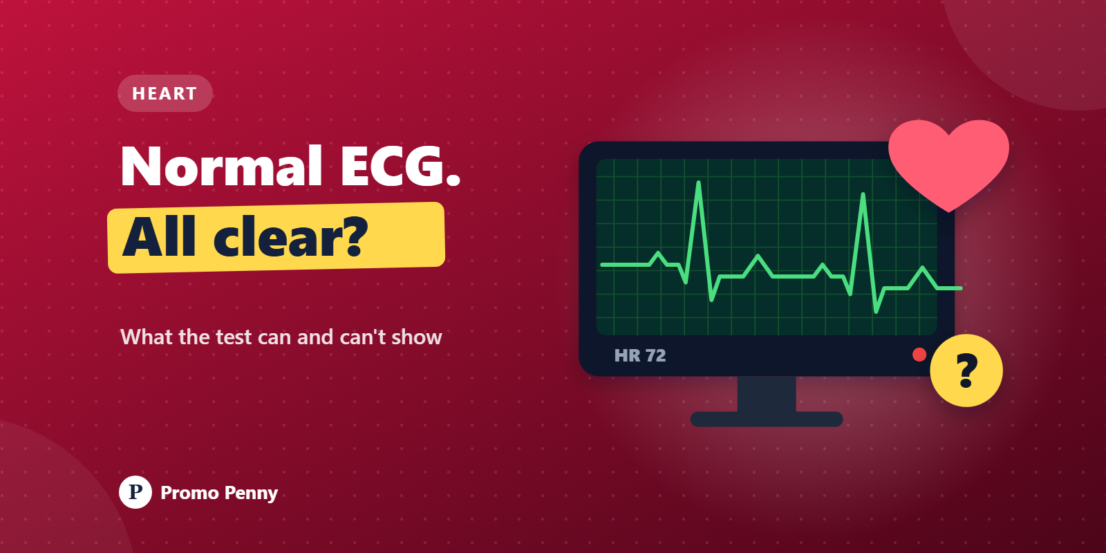
You lie on a narrow couch, a technician sticks cold electrodes on your chest, wrists and ankles, and in under a minute a strip of pink graph paper comes out of the machine. At the top, in small print, the machine has already written its opinion. Sometimes that opinion says normal. Sometimes it says something alarming.
The electrocardiogram, or ECG, is one of the most useful and most misunderstood tests in medicine. It is very good at some things and blind to others. Knowing which is which saves you from both false panic and false comfort.
Every heartbeat starts with a tiny electrical signal. It begins in a natural pacemaker at the top of the heart, spreads across the upper chambers (the atria), pauses briefly at a junction box, and then races through the lower chambers (the ventricles), which squeeze and pump blood out to the body.
An ECG picks up that electrical activity through the skin. Nothing goes into your body. The machine only listens. A standard 12-lead ECG uses ten electrodes, four on the limbs and six across the chest, and combines them to view the heart's electrical activity from twelve angles. The usual recording covers about ten seconds.
That last detail matters a lot, as you will see.
A normal beat on an ECG has a familiar shape, and each part has a name:
The grid helps too. At the standard paper speed, one big square equals 0.2 seconds. A quick rule of thumb for a regular rhythm is to divide 300 by the number of big squares between two spikes. For example, if the spikes are four big squares apart, the rate is about 300 / 4 = 75 beats a minute. Doctors use more careful methods, but it shows how the paper turns into numbers.
Within those ten seconds, an ECG can show a lot:
Here is the part that surprises people. A completely normal resting ECG does not rule out heart disease.
Take a hypothetical 50-year-old who gets breathless and tight-chested climbing stairs, but feels fine sitting down. He may have a significant narrowing in a coronary artery. At rest, his heart muscle gets just enough blood, so his resting ECG can look perfectly normal. The problem only shows up when the heart is made to work harder.
An ECG also misses a lot because of that short recording window:
So a normal ECG means something real: at that moment, the heart's electrical activity looked normal. It does not mean the arteries are clear or that your symptoms are nothing.
Depending on your symptoms and risk factors, a doctor may suggest one of these:
| Test | What it looks at | Typically used for |
|---|---|---|
| TMT (treadmill or exercise stress test) | ECG recorded while you walk on a treadmill at increasing effort | Chest discomfort or breathlessness on exertion, checking for reduced blood supply |
| Echocardiogram (echo) | Ultrasound pictures of the heart | Pumping strength, valves, heart muscle thickness, fluid around the heart |
| Holter monitor | ECG recorded continuously, often for 24 to 48 hours | Palpitations, dizzy spells, suspected intermittent rhythm problems |
| CT coronary angiography or coronary angiogram | Images of the coronary arteries themselves | When a narrowing is suspected and needs to be seen directly |
No single test answers every question. Which one comes next depends on what the doctor is trying to find out, which is why the same ECG can lead to very different next steps for two different people.
Most ECG machines print an automatic interpretation. It is handy, but it is a computer's first guess, not a diagnosis. These programs are known to flag harmless variations as abnormal and, less often, to miss real problems.
Phrases you might see that often turn out to be minor in context include sinus arrhythmia (a normal variation in rate with breathing, common in young people), sinus bradycardia (a slow rate, which can be normal in fit people and during sleep), nonspecific ST-T changes, and incomplete right bundle branch block. They can matter, but only a doctor looking at the actual tracing alongside your history can say whether they do.
A few more things that throw off readings: electrodes placed in the wrong spot, shivering or talking during the test, and poor skin contact. A repeat ECG often clears up a strange first result.
Smartwatch ECGs deserve a mention. Most record a single lead, which can be useful for spotting some irregular rhythms but is nowhere near a full 12-lead ECG, and cannot rule out a heart attack.
This article is general information, not medical advice. Please discuss your ECG report and any symptoms with a doctor.





























© 2026 Promo Penny. All rights reserved
Designed & Developed by IngeniousPro Team.